
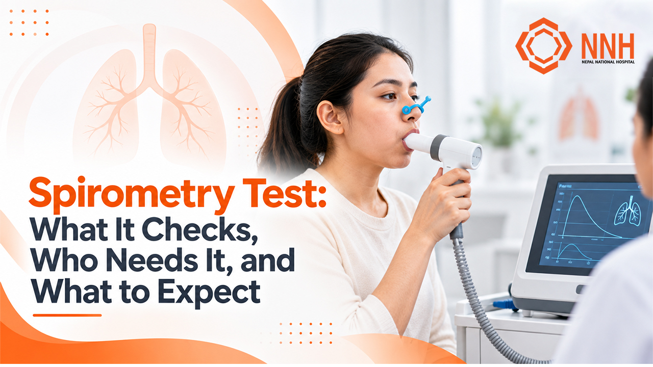
A spirometry test is the most common and foundational pulmonary function test (PFT) used in modern medicine. It measures how much air you can breathe into your lungs and how quickly and forcefully you can blow it out. By recording these airflow volumes over time, spirometry provides doctors with an objective, mechanical assessment of your bronchial health.
If you are experiencing persistent shortness of breath, a chronic cough, or unexplained wheezing or if your physician suspects conditions like asthma or Chronic Obstructive Pulmonary Disease (COPD) a spirometry test is the international standard used to confirm or rule out the diagnosis.
What Does a Spirometry Test Actually Check?
Spirometry assesses how well your airways function. When you breathe in, air travels through your trachea into branching bronchial tubes down to tiny air sacs (alveoli). If your airways are inflamed, swollen, constricted by muscle spasms, or obstructed by mucus, the speed at which air leaves your lungs decreases significantly.
The test records your breath using a precision device called a spirometer. The patient breathes through a filtered mouthpiece connected to a sensor that tracks airflow volume and velocity, generating two critical visual curves: a volume-time curve and a flow-volume loop.
   Normal Flow-Volume Curve vs. Obstructive Pattern
   Flow (L/s)
      ^
      |      /\  <-- Normal peak flow & smooth descent
      |     /  \
      |    /    \
      |   /  _---\_  <-- Obstructive "Scooped" curve (airway narrowing)
      |  / _-      \_
      +-------------------------> Volume (L)
The Core Lung Metrics: FVC, FEV1, and the Ratio
Your pulmonologist evaluates your lung function through three foundational numbers:
- Forced Vital Capacity (FVC):The total volume of air you can forcefully exhale in one continuous breath after filling your lungs to maximum capacity. Measured in liters, FVC indicates your overall functional lung volume.
- Forced Expiratory Volume in 1 Second (FEV1):The volume of air you can blast out of your lungs during the very first second of that forced exhalation. FEV1 is the single most important marker for detecting airway resistance and narrowing.
- The FEV1/FVC Ratio: This calculated fraction represents the proportion of your total lung capacity that you can exhale within the first second. In a healthy adult, this value generally ranges between 70% and 85% (or above the statistically defined Lower Limit of Normal, LLN). A significantly decreased ratio indicates that your airways are obstructed, trapping air inside the lungs.
According to the standardization guidelines published jointly by the American Thoracic Society (ATS) and European Respiratory Society (ERS), your measured numerical results are compared against predicted reference values matched to your age, biological sex, height, and ethnicity.
Pre- and Post-Bronchodilator Testing (Reversibility)
In many clinical evaluations, your doctor will order a reversibility test. This involves two distinct testing phases during the same visit:
- Baseline Spirometry: You perform the forced breathing maneuver without recent medication.
- Medication Inhalation: You inhale a fast-acting bronchodilator (such as 4 puffs of salbutamol/albuterol) through a spacer device.
- Repeat Spirometry: After waiting 15 minutes for the medicine to relax your bronchial smooth muscles, you repeat the breathing test.
If your FEV1 or FVC improves by more than 12% and at least 200 mL, the airway obstruction is classified as reversible a classic hallmark of bronchial asthma. If airflow limitation remains largely unchanged after the bronchodilator, it suggests fixed airway remodeling, typical of Chronic Obstructive Pulmonary Disease (COPD).
Who Needs a Spirometry Test?
Doctors do not order spirometry solely when severe lung disease is already obvious. It is a critical early-warning tool used across several clinical scenarios:
1. Investigating Persistent Respiratory Symptoms
You should consult a doctor regarding a spirometry test if you suffer from:
- Shortness of breath (dyspnea) during routine activities like climbing stairs or walking short distances.
- A chronic cough persisting for more than 4 to 8 weeks without a clear cause.
- Frequent chest tightness, especially at night or early in the morning.
- Wheezing a whistling or squeaking sound when you breathe out.
- Frequent, lingering chest infections that take weeks to resolve.
2. Diagnosing and Differentiating Asthma vs. COPD
While asthma and COPD share symptoms such as breathlessness and coughing, their underlying pathology, progression, and treatment approaches differ fundamentally.
- Asthma typically features variable, reversible airway inflammation often triggered by allergens, cold air, or exercise.
- COPD (including chronic bronchitis and emphysema) involves progressive, largely irreversible lung damage, frequently tied to long-term exposure to inhaled irritants.
Spirometry provides the objective physiological data required to distinguish between the two, preventing misdiagnosis and improper medication use.
3. Environmental and Occupational Risk Factors in Nepal
Urban centers across Nepal particularly the Kathmandu Valley frequently record elevated ambient levels of particulate matter (PM2.5 and PM10), seasonal road dust, and industrial emissions. Chronic exposure to these airborne particulates can trigger insidious airway inflammation.
Furthermore, individuals who grew up using indoor biomass fuel cookstoves (chulho) in poorly ventilated rural kitchens, as well as industrial workers in brick kilns, cement factories, textiles, and carpentry, carry an elevated risk of developing occupational lung diseases and chronic airflow obstruction. Spirometry detects small-airway impairment years before physical breathlessness becomes disabling.
4. Smoking and Tobacco Exposure
If you are a current or former cigarette or bidi smoker over the age of 35, proactive spirometry screening can identify early-stage COPD before irreversible damage occurs. The Global Initiative for Chronic Obstructive Lung Disease (GOLD) strongly advocates for spirometric confirmation in any individual with respiratory symptoms and a history of exposure to tobacco smoke or environmental biomass.
5. Pre-Operative and Clinical Monitoring
Spirometry is routinely conducted before major thoracic or upper abdominal surgeries to evaluate operative pulmonary risk. It is also used to track lung function changes in patients taking medications with potential pulmonary toxicity, or to assess the efficacy of newly initiated inhaler regimens.
How to Prepare for Your Spirometry Appointment
Accurate spirometry depends directly on patient preparation. Because medications can temporarily widen your airways and mask underlying obstruction, you may be instructed to withhold certain respiratory medicines prior to testing.
Crucial Safety Rule: Never discontinue any prescribed inhaler or heart medication without explicit confirmation from your prescribing doctor. If you are experiencing acute, severe breathlessness, use your rescue inhaler immediately and seek urgent medical care.
The table below summarizes standard medication withholding intervals established by the ATS/ERS technical standards:
| Medication Class | Common Examples | Typical Withholding Time |
| Short-Acting Beta-Agonists (SABA) | Salbutamol (Ventolin, Asthalin), Levosalbutamol | 4 to 6 hours before the test |
| Short-Acting Muscarinic Antagonists (SAMA) | Ipratropium bromide (Ipivent) | 12 hours before the test |
| Long-Acting Beta-Agonists (LABA) | Salmeterol, Formoterol | 24 hours before the test |
| Long-Acting Muscarinic Antagonists (LAMA) | Tiotropium, Glycopyrronium | 36 to 48 hours before the test |
| Ultra-Long-Acting Bronchodilators | Indacaterol, Vilanterol combinations | 48 hours before the test |
| Oral Bronchodilators / Theophylline | Theophylline, Aminophylline | 12 to 24 hours before the test |
Day-of-Test Instructions
To ensure your test maneuvers are valid and comfortable:
- Avoid heavy meals: Do not eat a large meal within 2 hours of your test; a distended stomach restricts diaphragmatic movement.
- Avoid vigorous exercise: Refrain from heavy workouts for at least 1 to 2 hours prior to testing.
- Do not smoke or vape: Avoid all tobacco, bidi, or electronic cigarettes for at least 1 hour before the test (or ideally 24 hours if assessing baseline lung health).
- Skip caffeine and alcohol: Avoid coffee, strong tea, energy drinks, and alcohol on the day of testing, as they can alter airway tone.
- Wear loose, comfortable clothing: Tight shirts, ties, corsets, or restrictive belts limit chest and abdominal expansion during maximal inspiration.
- Dentures: If you wear dentures, keep them in place unless they are loose; a secure mouth seal around the mouthpiece is essential to prevent air leaks.
Step-by-Step: What Happens During the Test?
A spirometry test requires active cooperation and physical exertion. You will work closely with a trained pulmonary function technician or nurse who will coach you through every breath.
+---------------------------------------------------------------------------------+
|                        THE SPIROMETRY CYCLE AT A GLANCE                         |
|                                                                                 |
|   1. REST               2. MAXIMAL INHALE      3. BLAST EXHALE   4. COMPLETE    |
|  [Breathe normally] -> [Fill lungs to max] -> [BLOW HARD & FAST] -> [Empty 6s] |
+---------------------------------------------------------------------------------+
1. Initial Setup and Measurements
- The technician will measure your exact height and weight (without shoes) and record your age. These parameters determine your predicted normal values.
- You will sit upright in a sturdy chair with your feet flat on the floor. Good posture ensures maximum chest excursion.
- A soft nose clip will be placed over your nostrils to prevent air from escaping through your nose.
2. The Forced Exhalation Maneuver
- You will place a clean, single-use filtered mouthpiece securely between your teeth and seal your lips tightly around it. Air must not leak from the corners of your mouth.
- You will breathe normally for a few cycles, then the technician will instruct you to inhale as deeply and fully as possible until your lungs are completely filled.
- Without hesitation, you will blast the air out as hard and fast as you possibly can.
- You must keep blowing continuously for at least 6 seconds (or 3 seconds for young children), even when you feel your lungs are entirely empty. The technician will actively cheer and urge you on: “Keep blowing! Push, push, push! Don’t stop!”
- Finally, you will take one rapid, complete breath back in through the mouthpiece to complete the loop.
3. Quality and Repeatability Criteria
To meet international clinical guidelines, a spirometry session cannot rely on a single breath:
- You must produce at least three acceptable maneuvers. An acceptable attempt has a clean, rapid start without coughing during the first second and lasts at least 6 continuous seconds.
- The results must be repeatable: your highest two FVC and FEV1 measurements must fall within 150 mL (0.15 L) of each other.
- Most sessions involve between 3 and 8 total blows to achieve acceptable, reproducible data.
The entire procedure typically takes 20 to 30 minutes (or 45 minutes if including a 15-minute bronchodilator reversibility pause).
Understanding Your Spirometry Results
Once testing is complete, the computerized spirometer prints a report featuring your raw numerical outputs, predicted percentages, and graphic flow curves. Your pulmonologist interprets these patterns to determine whether your ventilatory profile is normal, obstructive, restrictive, or mixed.
1. Normal Spirometry
- Both your FVC and FEV1 are equal to or greater than 80% of your predicted value.
- Your FEV_1/FVC ratio is normal (generally >0.70 or above your age-adjusted LLN).
- This indicates healthy lung volume and unobstructed airflow.
2. Obstructive Pattern
In an obstructive defect, air can enter the lungs normally, but narrowed airways impede how quickly it can be expelled.
- The Key Indicator: The FEV1/FVC ratio is below 0.70 (or below the LLN).
- FEV1 is disproportionately reduced compared to FVC.
- Common Causes: Asthma, COPD (chronic bronchitis/emphysema), bronchiectasis, and cystic fibrosis.
- Severity Grading: Based on post-bronchodilator FEV1 percentage:
- Mild: FEV1 >= 80% predicted
- Moderate: $50% <= FEV1 < 80% predicted
- Severe: 30% <= FEV1 < 50% predicted
- Very Severe: FEV1 < 30% predicted
3. Restrictive Pattern
In a restrictive defect, the total volume of air the lungs can hold is abnormally low, even though the airways themselves are not narrowed.
- The Key Indicator: The FEV1/FVC ratio is normal or elevated (>= 0.70), but total vital capacity (FVC) is reduced below 80% of predicted.
- Common Causes: Interstitial lung disease (pulmonary fibrosis), severe chest wall deformities (scoliosis), neuromuscular disorders, obesity hypoventilation, or pleural effusion.
- Note: Spirometry can strongly suggest restriction, but confirmation requires a formal Total Lung Capacity ($TLC$) measurement via plethysmography or gas dilution.
4. Comparison Table: Diagnostic Patterns
| Spirometry Metric | Normal Result | Obstructive Defect | Suspected Restrictive Defect |
| FEV1/FVC Ratio | Normal (>= 0.70) | Decreased (< 0.70) | Normal or High (0.70) |
| FEV1 | Normal (>= 80%) | Decreased | Decreased or Normal |
| FVC | Normal (>=80%) | Normal or Decreased | Decreased (< 80%) |
| Bronchodilator Response | None | Significant in Asthma; variable in COPD | Generally absent |
| Typical Clinical Examples | Healthy lungs | Asthma, COPD, Bronchiectasis | Pulmonary Fibrosis, Kyphoscoliosis |
Is Spirometry Safe? (Risks and Contraindications)
Spirometry is a safe, non-invasive procedure. However, the physical effort required to blast air out causes substantial pressure changes inside your chest, abdomen, skull, and eyes.
Common, Mild Sensations
During or immediately after the blows, it is completely normal to experience:
- Mild lightheadedness or dizziness (due to hyperventilation).
- Temporary breathlessness.
- A brief coughing reflex triggered by forceful air movement.
- Flushing or minor fatigue.
The technician will provide rest periods between efforts to ensure your comfort.
Contraindications: When the Test Must Be Postponed
Because the maneuver dramatically increases intrathoracic, intraocular, and intracranial pressures, spirometry should not be performed if you have any of the following conditions:
- Recent eye surgery (cataract or retinal surgery within the past 1 to 6 weeks).
- Recent thoracic or abdominal surgery (within the past 4 weeks).
- Recent myocardial infarction (heart attack) or unstable angina (within the past month).
- Known thoracic or abdominal aortic aneurysm.
- Recent pneumothorax (collapsed lung) within the past 2 to 4 weeks.
- Recent stroke, intracranial hemorrhage, or brain surgery.
- Active, untreated respiratory infections such as open pulmonary tuberculosis, COVID-19, or acute hemoptysis (coughing up blood).
Always disclose your complete medical, surgical, and cardiac history to your healthcare provider before booking the test.
Accessing Quality Spirometry Testing in Kathmandu
An accurate spirometry report requires two elements: modern, properly calibrated computerized equipment and an experienced pulmonary technician who knows how to coach patients to achieve maximal effort. Suboptimal effort, air leaks, or early exhalation termination produce falsely low readings that can lead to inappropriate medical treatments.
At Nepal National Hospital, our department of pulmonology and respiratory medicine offers standardized diagnostic pulmonary testing in a dedicated clinical environment. Whether you are dealing with seasonal coughs exacerbated by Kathmandu’s urban environment, seeking formal asthma management, or requiring pre-operative respiratory clearance, our clinical team provides comprehensive evaluations.
If your breathing feels labored or your daily life is limited by persistent coughing, schedule a consultation with a chest specialist to evaluate your lung health with objective, reliable diagnostics.
FAQs
Can I take a spirometry test if I currently have a common cold or chest infection?
It is generally recommended to postpone elective spirometry for 4 to 6 weeks after an acute viral upper respiratory tract infection, bronchitis, or pneumonia resolves. Acute airway inflammation causes temporary hyperresponsiveness and reduced airflow that can produce misleading results unreflective of your true baseline lung function.
Does the spirometry test hurt?
No, the test is entirely non-invasive and causes no physical pain. The primary sensation is the fatigue associated with blowing as hard and fast as possible several times in succession. You may experience momentary lightheadedness, which clears quickly after a few normal breaths.
Why does the technician raise their voice during the test?
Patients are often surprised when the technician vigorously shouts instructions like “Push, push, keep blowing!” This vocal coaching is a required, standardized element of pulmonary function testing. Research shows that enthusiastic verbal encouragement is essential to motivate patients to overcome the natural reflex to stop exhaling, ensuring the test captures true maximal capacity.
Can young children perform a spirometry test?
Spirometry can be successfully performed by most children starting between 5 and 6 years of age. It requires the child to understand and follow specific multi-step instructions. Specialized pediatric pulmonary labs often use interactive incentive animations (such as blowing out birthday candles or popping virtual balloons on a screen) to help younger patients maintain adequate exhalation effort.
What is the difference between spirometry and a peak flow meter?
A peak flow meter is a small, handheld plastic mechanical device that measures only your Peak Expiratory Flow (PEF) the maximum speed of air exiting your lungs in the first millisecond. It is primarily used at home by asthma patients to monitor day-to-day fluctuations. A spirometer is a precision medical diagnostic instrument that measures both volume (FVC) and speed (FEV1) over time, capable of formally differentiating between obstructive and restrictive lung diseases.